Neuroblastoma is a very common type of cancer in childhood. It is a tumor that forms in the nervous tissue and can be difficult to intervene because it is usually blood vessel wrapped which, if damaged, can end the child's life.
A 5-year-old boy suffered from it since he was one year old, in the belly. He was treated with chemotherapy to control the tumor and not go over, while they found the time and way to remove it. He was operated twice, but failed because he could not access the tumor without putting the child at risk. Finally, last month, as they explain in El Periódico, they achieved carry out the impossible intervention thanks to a 3D model of the child.
And it is that the new technologies, and especially the one of impression of materials in 3D, is proving a true route of solutions for the medicine, as they have told us in Xataka in some occasion.
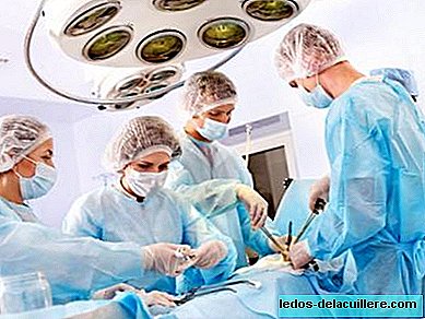
After chemotherapy and the two interventions doctors at the Sant Joan de Déu hospital in Barcelona could have thrown in the towel. It was an impossible operation. However, they did not give up and began to look for reliable ways to carry out the difficult intervention.
That was when they came up with the possibility of training. How to make possible something that seems impossible? Doing it many times, training, dominating what will be done at the critical moment. They contacted the CIM Foundation, a technological center of the Polytechnic University of Catalonia (UPC), and with their collaboration they managed to do models of the child's organs and tumor.
They made the impression in two different materials. On the one hand they made use of a resin to reproduce the blood vessels and organs and other softer and translucent material, of a consistency similar to the tumor, so that the surgeons tried to remove it without touching the vessels.
In addition, another model of the child's organs was made without the tumor to visualize the result they should achieve.
The model arrived 10 days before the operation and the surgeons had those days to work with her. They studied the best path, searched and found solutions and finally "operated" the model again and again. Thanks to this, on the day of the intervention, they knew perfectly well how they should access the tumor and how they should proceed to remove it. The benefit was not only knowing how to do it, but, being already trained, everything was faster and more reliable.
Now doctors say that after surgery the child will regain health and that will no longer require further interventions. Hopefully it will be like that.
The result has been so convincing that they have already commissioned the 3D model of two more patients, in order to address both interventions with all the guarantees.
Without a doubt, this is great news for everyone. We are talking about a tumor that a few months ago was impossible to intervene. The pity is that everything that has to do with research has less and less funds and more cuts, and perhaps this news is repeated less and less, or less frequently than what would be given if the researchers had more means.