Ultrasound is a technique that uses sound waves to show images of the baby inside the womb. Ultrasound, which would refer to the technique used, is also known as ultrasound, sonogram or ultrasonogram, and does not present the disadvantages of radiation (for example X-rays) to have an image of the inside of the body.
Ultrasound allows to obtain important information about the health of the fetus and the conditions present in the uterus, which is why it has been a great advance for the health of pregnancy and neonatal in those places where its use is extended.
The information obtained thanks to ultrasound in pregnancy It allows the doctor to plan the medical care of the pregnant woman and improve her quality of life, as well as detect certain abnormalities in the fetus or possible problems for childbirth.
Ultrasound early in pregnancy It is done to verify that the embryo implantation has been successful or to confirm that there is pregnancy.
Transvaginal ultrasound
The early utrasonido in pregnancy is usually through a transvaginal ultrasound, that is, performed through the vagina, and not externally, on the belly, as is usual in subsequent ultrasounds.
To perform a transvaginal ultrasound the specialist inserts a probe, called transducer, inside the vagina. The probe is covered with a condom and a gel. This probe sends sound waves that reflect body structures and a computer receives them and uses them to create an image that can be seen immediately on an image screen.
The specialist moves the probe into the area to observe the pelvic organs. The test is usually painless, although some women may experience mild discomfort from the probe pressure.
As we will see below, early ultrasound It is the first that is usually done to pregnant women to confirm pregnancy and obtain other information, and is usually done by this transvaginal ultrasound.
Transvaginal ultrasound is also used later in pregnancy to evaluate cases of threatened miscarriage, examine the placenta, find the cause of possible bleeding, monitor the growth of the embryo or the fetus in early pregnancy.
It can also be used as a guide during other tests (such as amniocentesis) and, towards the end of pregnancy, to see if the cervix is changing or opening when labor is beginning early.

When ultrasound is performed early in pregnancy
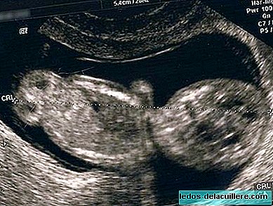
In the places where this prenatal control technique exists, transvaginal ultrasound is performed quite early in pregnancy, between weeks six and seven, to confirm if there is a pregnancy, diagnose a possible ectopic or molar pregnancy, and even detect heartbeats of the embryo. In addition, thanks to the previous data, it is possible to know if the pregnancy is simple or there is more than one fetus.
The embryo can be seen in a transvaginal ultrasound by six weeks of pregnancy (from the eighth week we will see the fetus). But even before pregnancy can be detected, since you can see the gestational sac. These dates are approximate, because depending on the accuracy of the equipment or the ability of the specialist to interpret the images, results can be obtained sooner or later.
Therefore in ultrasound early in pregnancy It is possible that the baby's heartbeat is not detected, but that does not mean that there is a problem if, for example, there is an error in the calculation of the pregnancy time and it is too early to detect it. In these cases, it is usually recommended to repeat the ultrasound one or two weeks later, or to perform an ultrasound with the Doppler technique.
Depending on the countries and medical centers in which the pregnant woman is monitored, ultrasound early in pregnancy will be performed sooner or later. There are even places where the first ultrasound is not done until 16 or 18 weeks, which could no longer be called “early”.
In any case, since ultrasound is a safe technique for both the mother and the baby as long as it is used properly by professionals, it would be advisable to do an early ultrasound to detect as soon as possible how the pregnancy started.
However, it should be noted that in low-risk women ultrasound is very useful to rule out problems, but it is not as effective in detecting them. And, in addition to not detecting some congenital defects, routine ultrasound can sometimes suggest the presence of a defect when in fact none exists, so it is recommended to do other studies to show if the baby is healthy or not.
Definitely, Early pregnancy ultrasound is a prenatal test that uses sound waves to capture the baby's first image inside the womb. This is the first ultrasound that will confirm the news and offer us more important data for the health of the fetus and ours.